Table of Contents
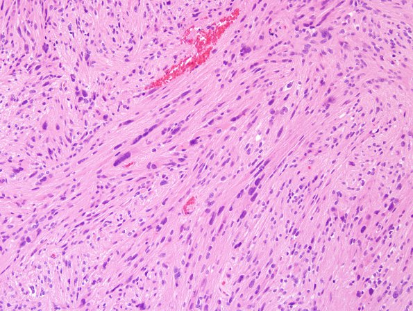
Washington University Experience | NEOPLASMS (GLIAL) | Ependymoma, tanycytic | 6A2 Ependymoma, tanycytic (Case 6) H&E 6
Microscopic examination of the generally well circumscribed lesion submitted shows a proliferation of bland cells with round to oval nuclei and inconspicuous nucleoli. Areas with the appearance of myxopapillary, clear cell and routine ependymoma are admixed. There are no mitoses, endothelial proliferation or areas of necrosis.

