Table of Contents
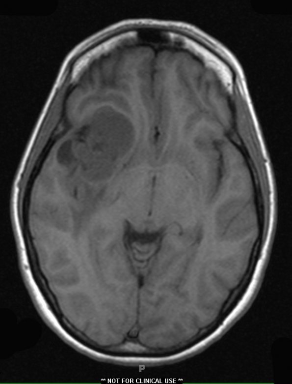
Washington University Experience | NEOPLASMS (GLIAL) | Ependymoma, tanycytic | 7A1 Ependymoma, (Case 7) T1 - Copy
Case 7 History ---- The patient is a 39-year-old woman who presented with headaches. MRI showed a mass in the right Sylvian fissure abutting the anterior right clinoid region measuring 4.0 x 2.7 cm. There is diffusion restriction associated with the mass, consistent with a cellular lesion. There is surrounding edema in the right frontal and temporal lobes. Operative procedure: Right frontotemporal craniotomy for tumor resection. 7A1-3 A mass with discrete borders is identified on this T1-weighted (7A1), T1-weighted with contrast (7A2) and T2-weighted with contrast (7A3) scans.

