Table of Contents
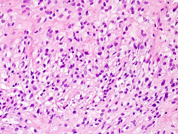
Washington University Experience | NEOPLASMS (GLIAL) | Ependymoma, tanycytic | 7B6 Ependymoma, (Case 7) H&E 1
Additionally, there are areas of focal clear cell ependymoma. Mitotic figures are identified at a rate of 2-3 per 10 high-powered fields. No microvascular proliferation or necrosis is seen.

