Table of Contents
Washington University Experience | NEOPLASMS (GLIAL) | Ependymoma - Electron Microscopy | 1B1 Ependymoma (Case 1) H&E 10
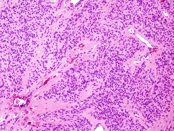
1B1-4 This is a neoplasm composed of small fibrillar to epithelioid cells with granular chromatin, forming perivascular pseudorosettes, ependyma lined cysts, and true ependymal rosettes. The nuclei are round to oval with prominent light to dark regions and scant cytoplasm. Mitotic figures are not easily identified. There is focal hemorrhage and scattered foci of non-pseudopalisading necrosis. (H&E)

