Table of Contents
Washington University Experience | NEOPLASMS (GLIAL) | Ependymoma - Electron Microscopy | 1C Ependymoma (Case 1) GFAP 1
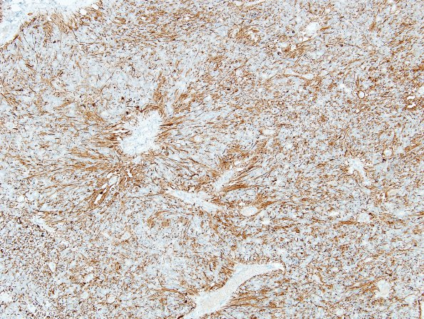
The tumor cells show diffuse positive staining for glial fibrillary acidic protein with GFAP rich fibrillary processes surrounding pseudo- and true rosettes. (GFAP IHC)

