Table of Contents
Washington University Experience | NEOPLASMS (GLIAL) | Ependymoma - Electron Microscopy | 2A1 Ependymoma (Case 2) FLAIR - Copy
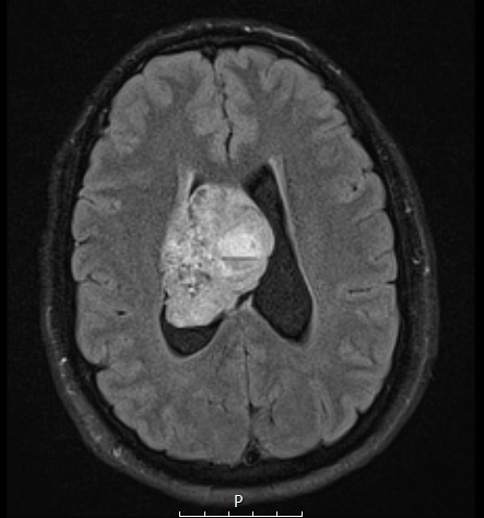
Case 2 History ---- The patient was a 23 year-old man who, after a roll-over motor vehicle accident, presented to an outside facility where head CT revealed a large heterogeneous right lateral ventricular mass. The patient experienced no loss of consciousness, headache, nausea, vomiting, or any neurologic signs and symptoms. ---- 2A1-3 Brain MRI shows a bubbly-appearing heterogenous mass with prominent cystic components within the right lateral ventricle. The lesion shows marked heterogenous contrast enhancement, the contrast enhancing portion measuring 4.5 x 4.3 x 6.2 cm. Angiography studies show that the tumor has vascularity comparable to that of normal brain, but with relatively persistent staining. Operative procedure: Craniotomy with tumor excision. ---- 2A1 A FLAIR image is hyperintense and shows multiple cysts.

