Table of Contents
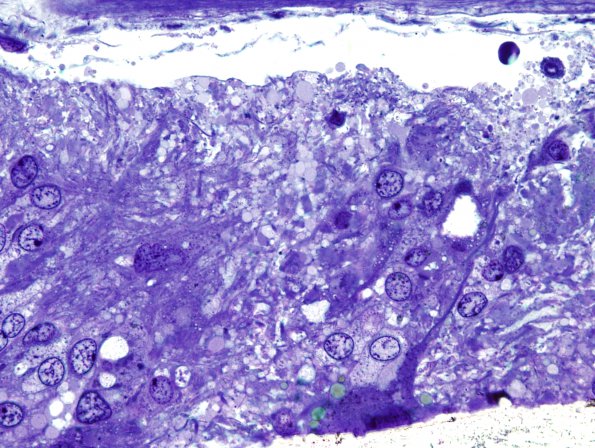
Washington University Experience | NEOPLASMS (GLIAL) | Ependymoma - Electron Microscopy | 2D3 Ependymoma (Case 2) 16
One-micron thick plastic embedded toluidine-blue stained images show individual tumor cells have a modest amount of relatively pale cytoplasm and extend thin tumor cell processes to form weak fascicles, and perivascular pseudorosettes. which are generally markedly hyalinized.

