Table of Contents
Washington University Experience | NEOPLASMS (GLIAL) | Ependymoma - Electron Microscopy | 3A1 Ependymoma, anaplastic (Case 3) 2.jpg
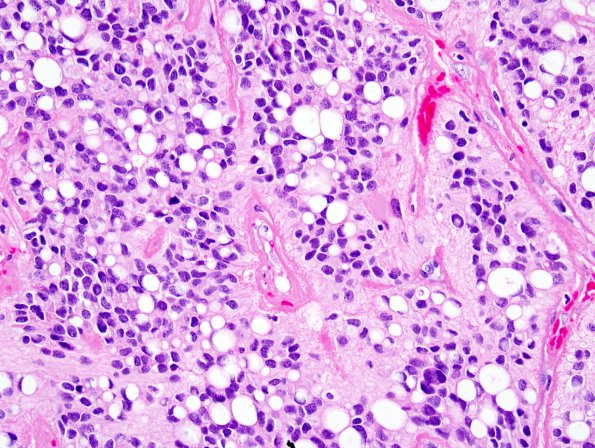
Case 3 History ---- The patient is a 29 year old woman who underwent resection of a primary brain tumor at the age of 15 months. She had several subsequent recurrences and treatment which included gamma knife radiation therapy. She recently presented with a heterogeneously enhancing right frontoparietal mass at the site of prior resections. ---- 3A1,2 The tumor cells have moderate nuclear pleomorphism, with the majority of nuclei being round to oval with mild hyperchromasia. The cells are associated with moderate quantities of clear to eosinophilic cytoplasm. Additionally, many of the cells have a signet-ring appearance with an eccentrically placed flattened nucleus displaced by a large clear cytoplasmic vacuole. In some areas, the tumor cells are arranged around blood vessels with a nuclear free zone, suggesting perivascular pseudorosettes. The mitotic index is high and there are many apoptotic nuclei. Foci of microvascular proliferation are present. Additionally, there are large areas of tumor necrosis, generally unassociated with nuclear pseudopalisading. In other areas, there is evidence of both radiation necrosis and radiation effects, including large zones of geographic, infarct-like necrosis associated with markedly hyalinized blood vessels and fibrinous exudates. Clusters of hemosiderin-laden macrophages suggest prior hemorrhage.

