Table of Contents
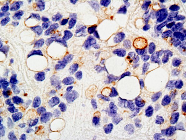
Washington University Experience | NEOPLASMS (GLIAL) | Ependymoma - Electron Microscopy | 3B2 Ependymoma, anaplastic (Case 3) EMA 2.jpg
The stain for epithelial membrane antigen (EMA) shows patchy immunoreactivity, including cytoplasmic dot-like positivity and membranous staining at the periphery of some of the clear cytoplasmic vacuoles in signet ring cells is also evident. ---- Not shown: A stain for GFAP shows patchy immunoreactivity and highlights perivascular pseudorosettes. A stain for cytokeratin 8/18 is negative.

