Table of Contents
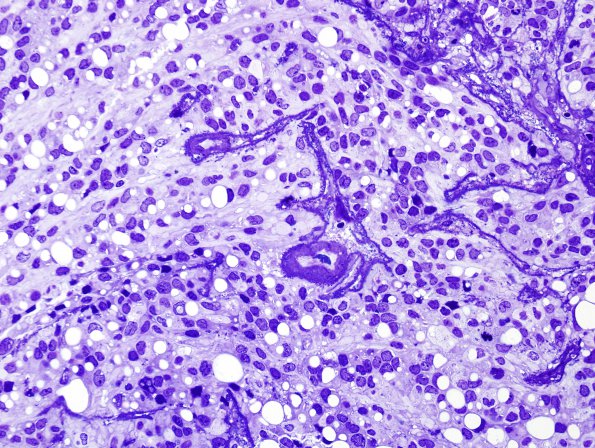
Washington University Experience | NEOPLASMS (GLIAL) | Ependymoma - Electron Microscopy | 3C1 Ependymoma, anaplastic (Case 3) Plastic 1 (2)
3C1,2 Electron microscopy was performed on submitted paraffin block material in order to further elucidate the tumor's pattern of differentiation. One-micron thick plastic embedded toluidine-blue stained images show individual tumor cells have associated small vacuoles, most of which appear to contain few cytosomes, specifically not aggregates of microvilli. Are these swollen lumina or the effect of extraction from paraffin or both?

