Table of Contents
Washington University Experience | NEOPLASMS (GLIAL) | Ependymoma - Electron Microscopy | 3D6 Case 3 Tumor_009 - Copy
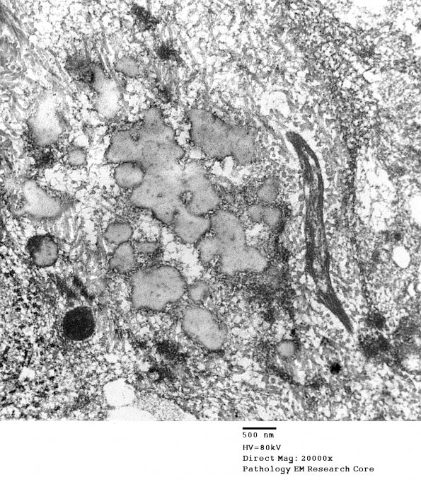
This amorphous material may be the matrix for eventual calcifications. (electron micrograph) ---- Comment: The morphologic, immunohistochemical, ultrastructural, and genetic features are most consistent with the diagnosis of anaplastic ependymoma, WHO grade III. The ultrastructural studies were particularly helpful for confirming this interpretation.

