Table of Contents
Washington University Experience | NEOPLASMS (GLIAL) | Ependymoma - Electron Microscopy | 4D1 Ependymoma, anaplastic (Case 4) Plastic 2 copy
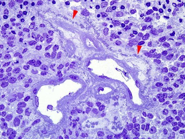
4D1-4 The vasculature of the tumor exhibits perivascular collections of amorphous material, some of which is a substrate for calcification (arrowheads). Tumor tissue was extracted from formalin-fixed paraffin-embedded tissue and embedding in plastic prior to preparation of one micron plastic sections; therefore, the preservation was not optimal. Nevertheless, one micron plastic sections showed perivascular punctate staining and occasional perivascular processes. (One-micron thick plastic embedded toluidine-blue stained images).

