Table of Contents
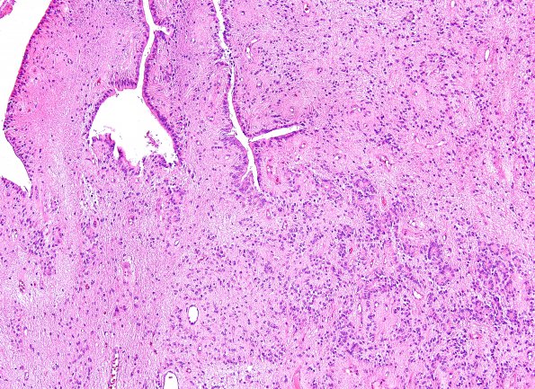
Washington University Experience | NEOPLASMS (GLIAL) | Ependymoma - Electron Microscopy | 6B1 Ependymoma, focal anaplasia (Case 6) H&E 1
6B1-4 Sections of this tumor show a glial neoplasm with prominent pseudo-rosettes, true rosettes and ependymal canals. It is moderately cellular, for most parts, and is composed of glial cells with regular round to ovoid nuclei. Most of the cells have indistinct cytoplasmic borders with few interspersed cells showing gemistocyte-like morphology. Mitoses are difficult to find in these portions of the tumor. Nonetheless, in some areas, the tumor appears hypercellular with relatively increased mitotic activity (~3/10HPF), nuclear pleomorphism and hyperchromasia. Large areas of geographic necrosis with patchy dystrophic calcification are present; in addition, rare foci of pseudopalisading necrosis are noted. Also, scattered minute foci of incipient 'glomeruloid' microvascular proliferation are present.

