Table of Contents
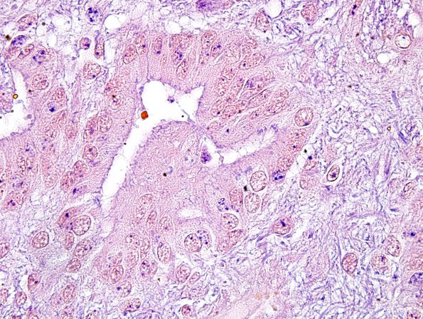
Washington University Experience | NEOPLASMS (GLIAL) | Ependymoma - Electron Microscopy | 6D3 Ependymoma, focal anaplasia (Case 6) PTAH 11
This PTAH stain also shows evidence of this terminal web (arrow). ---- Not shown:The tumor is immunoreactive for epithelial membrane antigen, CD99 and focal expression of mutant p53 protein, albeit with variable intensity. Further, lack of entrapped axons by a neurofilament stain is consistent with a solid growth pattern. A Ki-67 immunostain (performed on three different block) indicates a variable proliferation index, reaching ~9.5% in the hypercellular areas. ---- Comment: Presence of focal areas of hypercellularity (with relatively increased pleomorphism and mitoses), patchy incipient microvascular proliferation and rare foci of pseudopalisading necrosis are suggestive of focal anaplasia in an otherwise bland looking ependymoma and argue for a diagnosis of an ependymoma with focal anaplastic features (WHO grade 3).

