Table of Contents
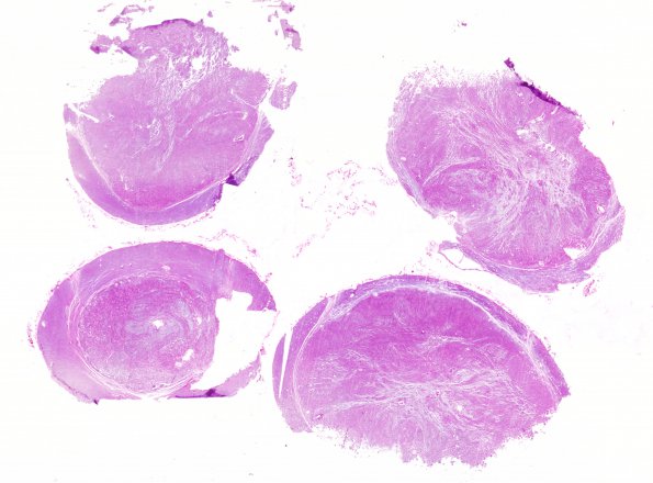
Washington University Experience | NEOPLASMS (GLIAL) | Ependymoma - Gross & Microscopic | 2A1 Ependymoma (Case 2) spinal H&E 2
Case 2 History ---- I almost discarded this case at a glance without a microscope thinking it was an example of toothpaste artifact. It is not. Rather, it is a fusiform tumor at the level of T-2 to T-5. Sections through this mass showed a sharp demarcation between the tumor and the rind of compressed but not invaded normal spinal cord. The tumor is well circumscribed, non-encapsulated, and consists of well differentiated spindled fibrillary cells with perivascular pseudorosettes, modest nuclear hyperchromatism and lacks mitotic activity. There is no vascular proliferation or necrosis. At the level of T3 and T4 the tumor entirely displaces white and grey matter leaving only small areas of anterior horns and white matter in both sides. At the most superior and inferior levels the tumor seems to replace only the posterior columns. ---- 2A1,2 H&E whole mounts of several spinal cord segments of which the thoracic cord represents the epicenter of the tumor and the tumor tapers superior and inferior in the cord. (H&E)

