Table of Contents
Washington University Experience | NEOPLASMS (GLIAL) | Ependymoma - Gross & Microscopic | 3A2 Ependymoma, anaplastic (Case 3) A1
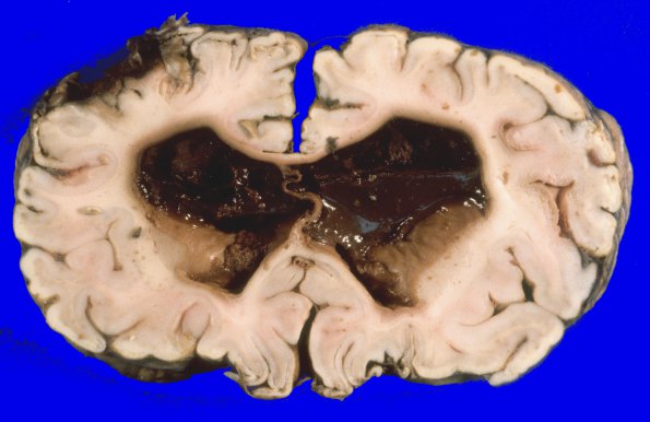
Serial coronal sections of the cerebral hemispheres at 2 cm intervals reveal marked distention of the lateral ventricles, which are filled by blood, mucinous material and fragments of friable soft tissue that have the same appearance and consistency as the main tumor mass. The cortex is of average and uniform thickness throughout. The convolutional and central white matter are markedly attenuated.

