Table of Contents
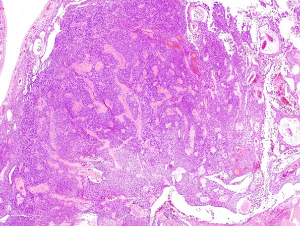
Washington University Experience | NEOPLASMS (GLIAL) | Ependymoma - Gross & Microscopic | 5C3 Ependymoma (Case 5), mimicking MPE, L2, H&E 6
5C3-5 A closeup of the solid areas shows a cellular tumor resembling a typical ependymoma rather than myxopapillary ependymoma. This tumor consist of a moderately cellular neoplasm arranged in a prominent pseudopapillary pattern. Many of these structures have hyalinized blood vessels in their center and are sheathed in ependymal cells as extensive canals. The tumor cells are bland, short spindled to epithelioid, have eosinophilic cytoplasm and contain nuclei with dispersed chromatin pattern. There is mild to moderate pleomorphism and scattered mitoses; the count reaching ~5/10HPF in some solid areas. A tiny area of infarct-like necrosis is also seen.

