Table of Contents
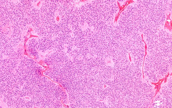
Washington University Experience | NEOPLASMS (GLIAL) | Ependymoma - Gross & Microscopic | 6B1 Ependymoma, cervical spinal cord (Case 6) H&E 10X
6B1,2 Histological sections of the spinal cord tumor show a well-circumscribed neoplasm composed of cells with uniformly sized round-to-oval nuclei, speckled chromatin, small eosinophilic nucleoli, eosinophilic cytoplasm, long thin bipolar cell processes, and indistinct cell borders. Pseudorosette formation is widespread; occasional true rosettes are present. The peripheral edge of the tumor, and a few clefts within the tumor tissue, are lined by tumor cells arranged to resemble a pseudostratified columnar epithelium with an underlying anucleate zone of cell processes. Focally, the epithelium exhibits intact cilia. Mitotic figures are not widespread, generally appearing at a density of <1/10HPF. Geographic necrosis is evident, without notable pseudopalisading by tumor cells. Immunohistochemically stained sections show nuclear reactivity for the proliferation marker Ki67 (MIB-1 antibody) at a density that focally ranges up to 2% of tumor cells. This histological/immunohistochemical pattern is consistent with ependymoma, WHO grade 2.

