Table of Contents
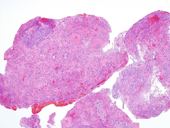
Washington University Experience | NEOPLASMS (GLIAL) | Ependymoma - Microscopic | 11B1 Ependymoma, anaplastic (Case 11) H&E 1
11B1-4 Sections consist of an ependymoma with prominent perivascular pseudorosettes and focal dystrophic calcification. In most areas, the tumor is only moderately cellular; however, the tumor is quite hypercellular in some areas with increase in nuclear-cytoplasmic ratio, hyperchromasia, brisk mitotic activity (~ 9/10 HPF), rare foci of early microvascular proliferation and areas of necrosis.

