Table of Contents
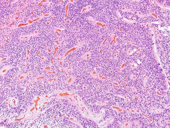
Washington University Experience | NEOPLASMS (GLIAL) | Ependymoma - Microscopic | 13A1 Ependymoma, anaplastic (Case 13) H&E 11
Case 13 History ---- The patient was a 2 year old boy with a two week history of dizziness and occasional balance problems. Head CT showed a 4.5 x 4.4 cm mass lesion within the posterior fossa. Operative procedure: Suboccipital craniotomy with resection of posterior fossa tumor. ---- 13A1,2 This is a hypercellular glial neoplasm with prominent perivascular pseudorosette formation. The neoplasm consists of cells with ovoid to round nuclei with irregular nuclear contours and stippled chromatin. Portions of the specimen show areas with lack of cohesion containing neoplastic cells with plump eosinophilic cell bodies and eccentrically located nuclei, suggestive of a 'rhabdoid' morphology. Endothelial hyperplasia is not present. Focal necrosis is observed. Large swaths of necrosis and pseudopalisading necrosis are not present. Neither true 'ependymal' rosettes nor Homer-Wright rosettes are present. Mitotic figures number up to 11 mitoses/10HPF.

