Table of Contents
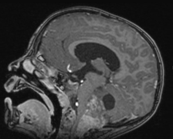
Washington University Experience | NEOPLASMS (GLIAL) | Ependymoma - Microscopic | 14A1 Ependymoma, Anaplastic (Case 14) T1 MPRAGE W 2 - Copy
Case 14 History ---- The patient was a 4 year old boy with headaches, vomiting and a wide based gait. Head CT and MRI of the brain show a large heterogeneously enhancing posterior fossa mass with areas of coarse calcification along with dilatation of the lateral, third, and fourth ventricles. Operative procedure: Posterior fossa craniotomy, C1 and partial C2 laminectomy with resection of tumor. ---- 14A1,2 The mass measures 6.3 x 3.6 x 4.2 cm and compresses the pons, medulla, and posterior aspect of the upper cervical spinal cord as shown in these contrast administered T1-weighted (14A1) and T2-weighted (14A2) scans.

