Table of Contents
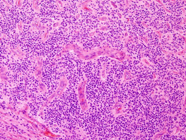
Washington University Experience | NEOPLASMS (GLIAL) | Ependymoma - Microscopic | 15B3 Ependymoma, anaplastic (Case 15) H&E 9.jpg
15B3,4 H&E stains show a hypercellular high grade ependymal neoplasm with frequent mitoses, microvascular proliferation, and patchy necrosis. The tumor has a solid growth pattern and there are perivascular pseudorosettes. True rosettes and true papillary/clefts are not identified. The tumor cells are tightly packed without distinct cell-cell borders and the nuclei are in a background of fibrillary neuropil. The tumor nuclei show modest pleomorphism, are round to slightly elongated, and largely have finely speckled hyperchromatic chromatin. Mitotic figures are frequent (>50 mitoses/10 HPF).

