Table of Contents
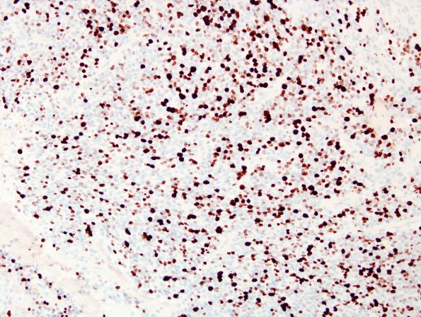
Washington University Experience | NEOPLASMS (GLIAL) | Ependymoma - Microscopic | 15E Ependymoma, anaplastic (Case 15) Ki67 2.jpg
The proliferation marker Ki67 stains an elevated subset of tumor cell nuclei at variable density, ranging focally up to approximately 44%. ---- Not shown:
Synaptophysin also demonstrates some weak dot-like positivity within the tumor cells. A subset of the tumor nuclei are positive for Olig2 and p53. The tumor is negative for cytokeratin (CK) and neurofilament (NF), the latter of which highlights the solid growth pattern of the tumor. ---- Comment: The overall histomorphologic and immunophenotypic characteristics are those of an ependymoma, WHO Grade 3.

