Table of Contents
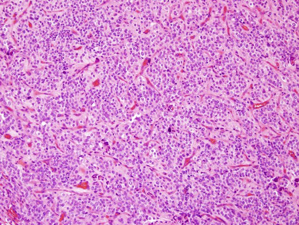
Washington University Experience | NEOPLASMS (GLIAL) | Ependymoma - Microscopic | 16B1 Ependymoma, anaplastic (Case 16) H&E 7.jpg
16B1-3 This is a cellular neoplasm comprised of small to medium sized cells with a high nuclear-to-cytoplasmic ratio. The tumor cells have round nuclei with vesicular chromatin and occasional prominent nucleoli and moderate amounts of amphophilic cytoplasm. There are scattered areas that show pseudorosettes; however, the majority of sections show a relative paucity of these structures. Mitoses were readily identified in multiple sections. The most mitotically active region showed 7 mitoses/ 10 HPF. There is focally prominent microvascular proliferation.

