Table of Contents
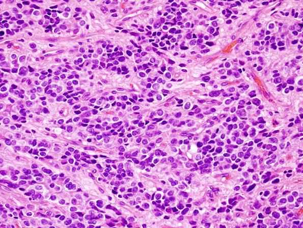
Washington University Experience | NEOPLASMS (GLIAL) | Ependymoma - Microscopic | 16B3 Ependymoma, anaplastic (Case 16) H&E 5.jpg
There are scattered areas that show pseudorosettes; however, the majority of sections show a relative paucity of these structures. Mitoses were readily identified in multiple sections. The most mitotically active region showed 7 mitoses/ 10 HPF. There is focally prominent microvascular proliferation.

