Table of Contents
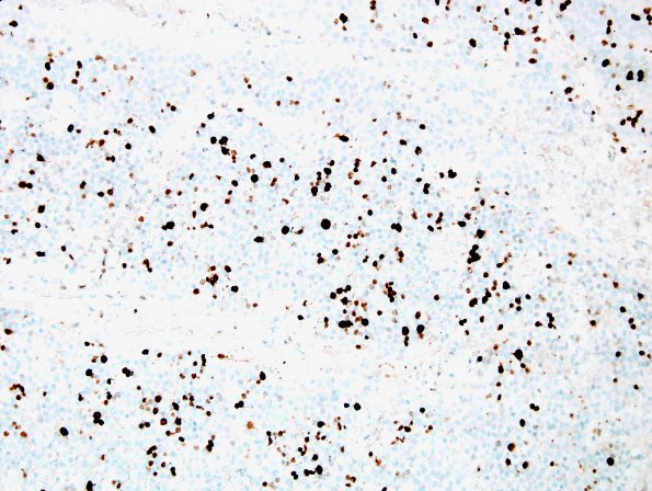
Washington University Experience | NEOPLASMS (GLIAL) | Ependymoma - Microscopic | 16F Ependymoma, anaplastic (Case 16) Ki67 1.jpg
Ki- 67 showed a proliferative index of 22.8%. ---- Not shown: CD34 highlights the vascular network but is negative in the tumor cells. NeuN demonstrates rare entrapped neurons. Synaptophysin shows a weak dot-like staining pattern with the tumor cells. ----
These histologic and immunohistochemical findings are consistent with a diagnosis of ependymoma, WHO Grade 3.

