Table of Contents
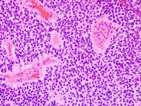
Washington University Experience | NEOPLASMS (GLIAL) | Ependymoma - Microscopic | 17A2 Ependymoma, anaplastic (Case 17) H&E 2
Sections of the resected parieto-occipital lesion show a neoplasm with multifocal tumor necrosis, including areas suspicious for pseudopalisading necrosis, and microvascular proliferation. Tumor cells appear in highly cellular sheets separated by hyalinized vessels with a suggestion of perivascular sparing. The growth pattern is largely solid with a sharp junction between the tumor and adjacent brain, although an infiltrative pattern is also present focally. Cytologically, neoplastic cells show high nucleus to cytoplasm ratio, moderate pleomorphism and evenly distributed chromatin without prominent nucleoli. Mitoses are easily identified.

