Table of Contents
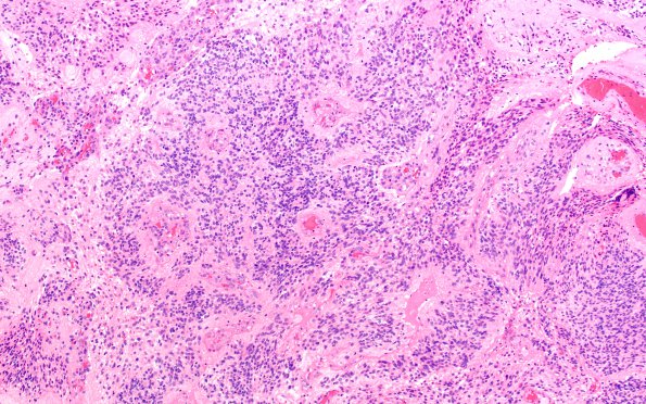
Washington University Experience | NEOPLASMS (GLIAL) | Ependymoma - Microscopic | 18B1 Ependymoma, anaplastic (Case 18) H&E 1
18B1-4 Sections show a glial neoplasm. Tumor cells have round to oval, slightly irregular nuclei with moderate amounts of eosinophilic cytoplasm. The tumor is arranged in prominent perivascular pseudorosettes. There is increased cellularity, necrosis, and microvascular proliferation. Mitotic activity is increased (focally up to 6/10 HPF). Frequent karyorrhectic debris is seen.

