Table of Contents
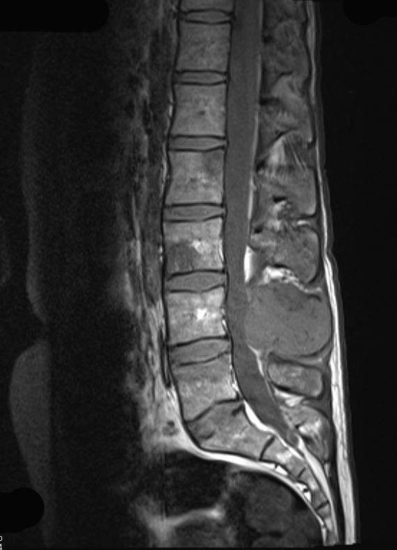
Washington University Experience | NEOPLASMS (GLIAL) | Ependymoma - Microscopic | 19A1 Ependymoma, anaplastic, metastatic WHO III (Case 19) T1 - Copy - Copy - Copy
Case 19 History ---- The patient was a 25-year-old woman with a history of left occipital ependymoma. The patient's original resection was performed in 2009 and a second resection was performed in 2012 at an outside hospital. More recently, the patient underwent a third resection of a tumor in the left occipital region which was shown to be ependymoma, WHO grade 3. FISH was negative for EGFR amplification. Prior to the third resection, the patient had Gamma knife radiotherapy. She then presented in September of 2014 with residual enhancing tissue in the anterior left occipital area, involving the dura and sagittal sinus. Additionally, there were osseous lesions at multiple levels in the thoracic and lumbar spine, felt to potentially represent metastatic disease. ---- Of note, she had a large infiltrating mass within the soft tissues and posterior elements of L4, as well as L3 and L5 which represents the tumor examined in the case shown here. Operative procedure: Resection of lumbar mass. 19A1-4 ---- 19A1,2 show the tumor in the dorsal spine compressing the spinal cord in this T1-weighted scan without (19A1) and with (19A2) administered contrast resulting in enhancement.

