Table of Contents
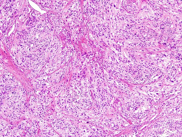
Washington University Experience | NEOPLASMS (GLIAL) | Ependymoma - Microscopic | 19B1 Ependymoma, anaplastic, metastatic WHO III (Case 19) H&E 6.jpg
19B1-5 These images are of a malignant tumor characterized by extensive fibrocollagenous tissue involved by irregular cords and nests of highly atypical and pleomorphic cells. Many of the cells show elongated eosinophilic processes with a syncytial growth pattern. Other cells appear more epithelioid. Nuclear atypia is marked, and there are scattered multinucleated forms seen. Many of the nests show central necrosis. Mitotic activity is noted, counted at a rate of approximately 2-3/10 HPF. The tumor shows involvement of the adjacent bone. Additionally, there appear to be fragments of collagenous ligament material also involved by tumor.

