Table of Contents
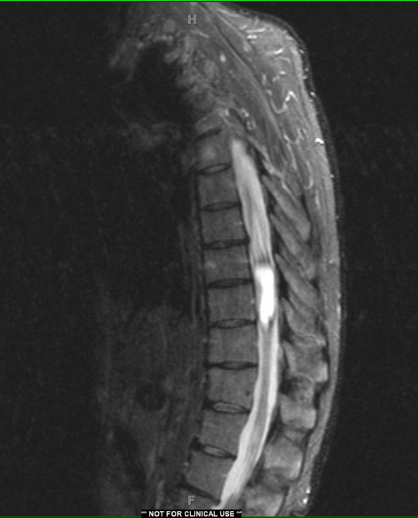
Washington University Experience | NEOPLASMS (GLIAL) | Ependymoma - Microscopic | 1A1 Ependymoma (Case 1) STIR no contrast - Copy
Case 1 History ---- The patient was a 47-year-old male with a long-standing history of back pain and scoliosis. Spine MRI showed a T2
hyperintense intramedullary cystic mass with peripheral intrinsic T1 hyperintensity and enhancement. Operative procedure: T8-T9 laminectomy, partial T7 through T10 laminectomy with decompression; resection of intradural intramedullary thoracic spinal cord lesion. ---- 1A1 This STIR (T1-weighted without contrast) image shows a hyperintense intramural mass.

