Table of Contents
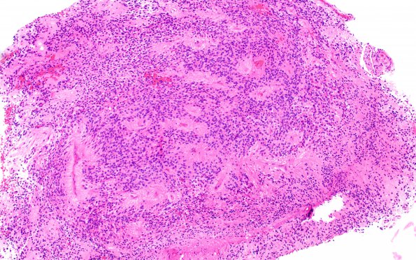
Washington University Experience | NEOPLASMS (GLIAL) | Ependymoma - Microscopic | 1B1 Ependymoma (Case 1) H&E 2
1B1,2 H&E shows tumor cells have round to oval, relatively regular nuclei. Focal true rosettes and perivascular pseudorosettes are seen. There is no significant mitotic activity. Vascular proliferation or necrosis are not identified.

