Table of Contents
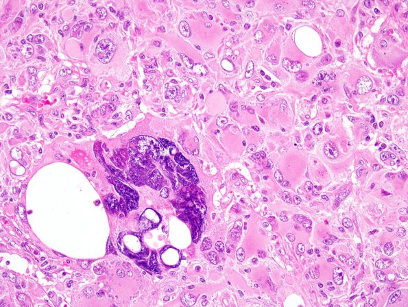
Washington University Experience | NEOPLASMS (GLIAL) | Ependymoma - Microscopic | 21A2 Ependymoma, giant cell (Case 21) H&E 1.jpg
This is a highly cellular relatively solid appearing neoplasm arranged in nests surrounded by fibrovascular septi. There is moderate to marked nuclear pleomorphism, as well as foci of microvascular proliferation and necrosis, focally associated with pseudopalisading. Multinucleated giant cells are seen in some areas. The mitotic index is brisk and there are frequent atypical mitoses. The tumor also extends to an ependymal surface in some areas. The interface with adjacent brain appears sharp. The tumor has a fibrillary appearance in some areas and some tumor cells resemble gemistocytes. Perivascular pseudorosettes are not a prominent feature on routine histology.

