Table of Contents
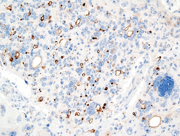
Washington University Experience | NEOPLASMS (GLIAL) | Ependymoma - Microscopic | 21D Ependymoma, giant cell (Case 21) EMA 1.jpg
EMA immunohistochemistry shows a similar pattern to D2-40. ----Not shown: Rare tumor cells show immunoreactivities for neurofilament protein and actin. The former stain also highlights axons at the periphery of the tumor only, consistent with a solid growth pattern. The tumor cells are negative for keratin, CK7, and CK20. Tumor nuclei showed retained expression for INI-1. Patchy dot-like positivity is seen in tumor cells with CD99 stain. The tumor is negative for desmin. ----
Comment: The morphologic and immunohistochemical features are consistent with the diagnosis of ependymoma with giant cell features, WHO grade 3. Although none of the stains are entirely specific for ependymal differentiation, the dot-like positivity with EMA, CD99, and D2-40 all support the diagnosis of ependymoma. Additionally, the solid, rather than infiltrative growth pattern of this tumor fits well:

