Table of Contents
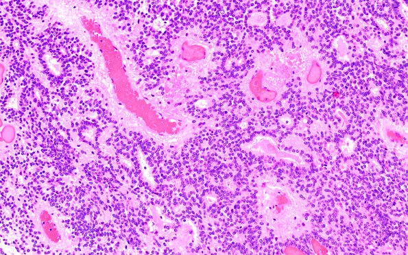
Washington University Experience | NEOPLASMS (GLIAL) | Ependymoma - Microscopic | 22B Ependymoma, WHO II (Case 22) H&E 3
Microscopy shows a neoplasm with atypical round to oval nuclei with speckled chromatin forming perivascular pseudorosettes and numerous true rosettes. Geographic necrosis is identified. Rosenthal fibers are identified in the reactive brain parenchyma adjacent to the lesion.

