Table of Contents
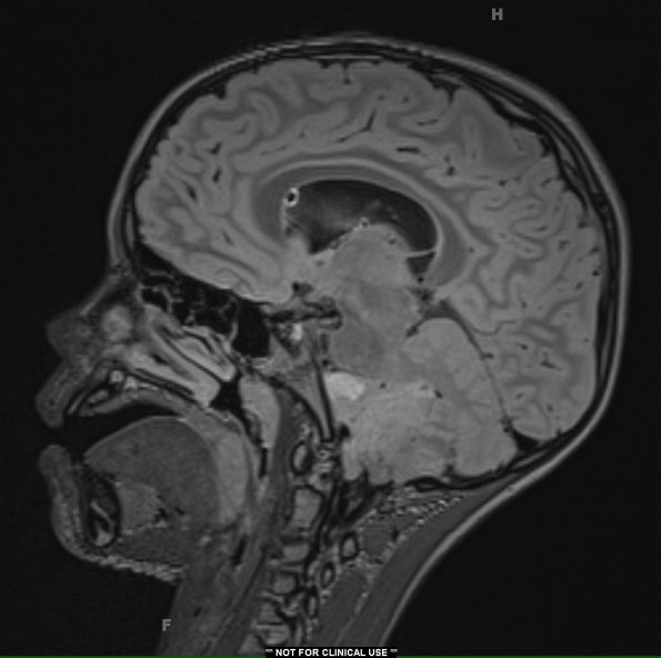
Washington University Experience | NEOPLASMS (GLIAL) | Ependymoma - Microscopic | 23A Ependymoma, focal anaplasia (WHO III) (Case 23) FLAIR 1 - Copy
Case 23 History ---- The patient was a 6-year-old boy with worsening headache and vomiting for the past week with positive bilateral nystagmus and left cranial nerve XII palsy. Brain imaging studies showed a predominantly cystic, enhancing extra-axial mass involving the left cerebellopontine angle. which exerts mass effect upon the pons and cerebellum with extension into the 4th ventricle. There is also mild ventriculomegaly with prominent cavum septum pellucidum. He underwent EVD placement with fenestration of septum pellucidum cyst and biopsy on 9/29. He subsequently underwent a posterior fossa craniotomy for tumor resection on 9/30 (this specimen). Operative procedure: Posterior fossa craniotomy for tumor resection. ---- 23A MRI studies (FLAIR shown here) showed a predominantly cystic, enhancing extra-axial mass involving the left cerebellopontine angle. which exerts mass effect upon the pons and cerebellum with extension into the 4th ventricle.

