Table of Contents
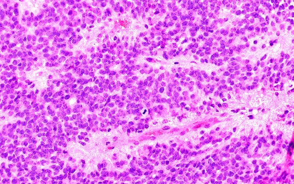
Washington University Experience | NEOPLASMS (GLIAL) | Ependymoma - Microscopic | 23B Ependymoma, focal anaplasia (WHO III) (Case 23) H&E 1
The tumor has a solid appearance and is composed of a monomorphic population of cells with round nuclei featuring crisp nuclear borders, inconspicuous nucleoli and speckled chromatin. The cells generally have eosinophilic cytoplasm, however there are areas with clear cell change, and they contain fibrillary cytoplasmic processes that regularly converge to form both perivascular rosettes and true rosettes. Large ependymal canals are present. Scattered throughout the tumor are a few nodules reminiscent of subependymoma. Mitotic figures are brisk in some areas. Focal areas of necrosis are seen, however there is no evidence of microvascular proliferation.

