Table of Contents
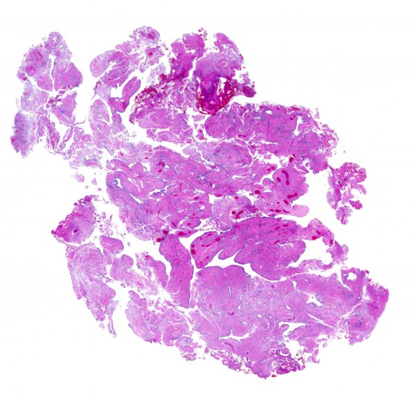
Washington University Experience | NEOPLASMS (GLIAL) | Ependymoma - Microscopic | 24A1 Ependymoma, WHO II (Case 24) H&E WM
Case 24 History ---- The patient was a 44 year old man with a spinal cord tumor extending from levels C4 to C7. Operative procedure: Excision ---- 24A1-4 H&E show multiple fragments of a solid growing ependymal neoplasm with lobular architecture and rounded pushing borders. The lobules have almost a papillary-like architecture containing many medium to large vessels. Surrounding these vessels is largely hypocellular dense fibrillary tissue. Some vessels do have perivascular pseudorosettes. These glial cells are relatively small and without distinct cell borders. Their nuclei are round to slightly elongated, slightly irregular, and have hyperchromatic chromatin. The epithelioid cells at are mostly arranged in a monolayer and range from columnar to flattened cuboidal in morphology. They have eosinophilic cytoplasm and nuclei that resemble those found in the glial component of the tumor. Definitive mitotic figures are not readily identified. There is no necrosis or microvascular proliferation identified in the tissue examined. Also, there are no true papillary structures or areas of myxoid material. Scattered chronic inflammatory cells are present throughout the tumor.

