Table of Contents
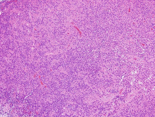
Washington University Experience | NEOPLASMS (GLIAL) | Ependymoma - Microscopic | 25B1 Ependymoma, focal anaplasia, Grade III (Case 25) H&E 1.jpg
25B1,2 Sections show a tumor with multiple areas differing in appearance and cell density. Some areas show a specimen with slightly increased cellularity and composed primarily of cells with long spindled cytoplasm, and oval to elongated nuclei with finely stippled chromatin and inconspicuous nucleoli. Other areas have a diffuse hypercellular appearance. (H&E)

