Table of Contents
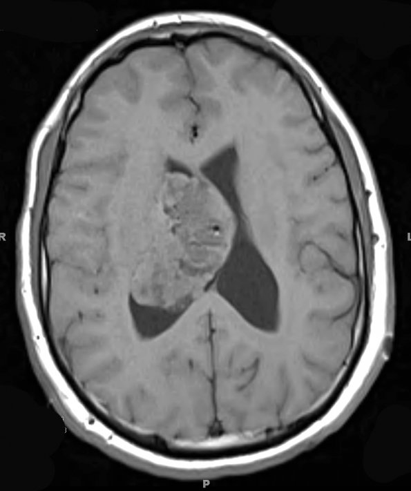
Washington University Experience | NEOPLASMS (GLIAL) | Ependymoma - Microscopic | 3A1 Ependymoma (Case 3) T1 4 - Copy
Case 3 History ---- The patient was a 23 year-old man who, after a roll-over motor vehicle accident, presented to an outside facility where computed tomography of the head revealed a large heterogeneous right lateral ventricular mass. Per clinical records, the patient experienced no loss of consciousness, headache, nausea, vomiting, or any neurologic signs and symptoms. MRI showed an intraventricular mass. Angiography studies show that the tumor had vascularity comparable to that of normal brain, but with relatively persistent staining. Operative procedure: Craniotomy with tumor excision. ---- 3A1,2 MRI showed a bubbly-appearing heterogenous mass with prominent cystic components and fluid/fluid levels within the right lateral ventricle. The lesion is isointense with this T1-weighted scan (3A1) and shows marked heterogenous contrast enhancement (3A2); the contrast enhancing portion measured 4.5 x 4.3 x 6.2 cm and was well demarcated from the adjacent brain parenchyma.

