Table of Contents
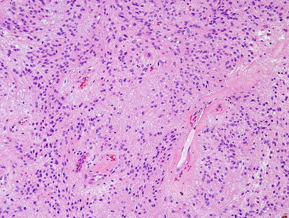
Washington University Experience | NEOPLASMS (GLIAL) | Ependymoma - Microscopic | 3B1pendymoma (Case 3) H&E 1
3B1,2 Histological sections of the tumor show a mildly hypercellular heterogenous neoplasm, most of which showing an irregular alternating distribution of tumor cell nuclei and an anuclear tumor neuropil. Individual tumor cells have a modest amount of relatively pale cytoplasm and extend thin tumor cell processes to form weak fascicles and perivascular pseudorosettes surrounding intratumor vessels. Although some nuclear pleomorphism is observed, the neoplastic cells generally have round to oval nuclei with relatively open speckled chromatin, crisp nuclear borders, and inconspicuous nucleoli.

