Table of Contents
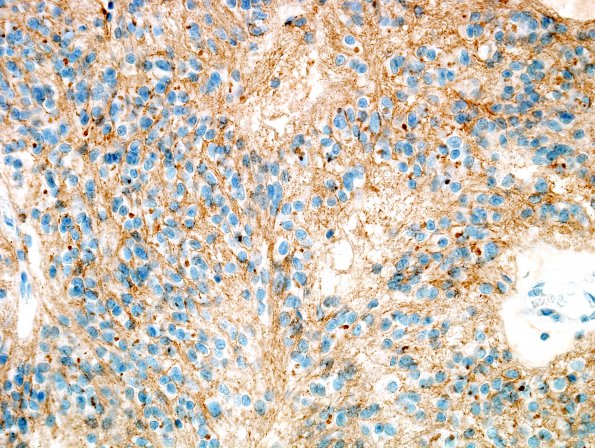
Washington University Experience | NEOPLASMS (GLIAL) | Ependymoma - Microscopic | 3D2 Ependymoma (Case 3) CD99 1
Reactivity for CD99 is moderate and widespread, and generally membranous, although it also appears focally in more intense paranuclear dots. ---- Not shown: The neoplastic cells show no reactivity for neuronal marker Neu-N. Nuclear reactivity for proliferation marker Ki-67 (MIB-1 antibody) stains tumor cell nuclei at low but variable density, quantified focally at 1.8%. Reactivity for Melan-A appears in a subset of tumor cells in the area that appears pigmented. ---- Comment: This histological/immunohistochemical pattern is that of ependymoma, WHO grade 2. Foci of infarct-like necrosis are not uncommon among these tumors and do not portend a more aggressive clinical course.

