Table of Contents
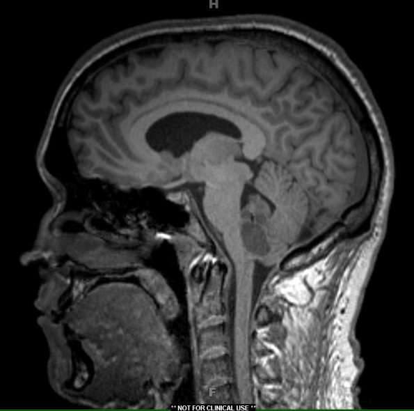
Washington University Experience | NEOPLASMS (GLIAL) | Ependymoma - Microscopic | 5A Ependymoma, WHO II (Case 5) T1 MPRAGE 1 - Copy
Case 5 History ---- The patient was a 30-year-old man with a 4.2 cm fourth ventricle multicystic mass with nodular contrast enhancement. There is mild enlargement of the lateral ventricles and the third ventricle. The radiographic differential diagnosis included ependymoma, choroid plexus papilloma, medulloblastoma and pilocytic astrocytoma. Operative procedure: Suboccipital craniectomy and excision of tumor. ---- 5A The ventricular neoplasm is hypo- & iso-intense in this T1-weighted image.

