Table of Contents
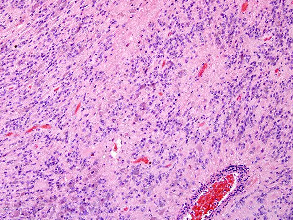
Washington University Experience | NEOPLASMS (GLIAL) | Ependymoma - Microscopic | 5B1 Ependymoma, WHO II (Case 5) H&E 1.jpg
Sections of the resected fourth ventricle tumor show a neoplasm arranged in streaming sheets of varying degrees of hypercellularity, frequent perivascular pseudorosettes and vascular hyalinization. Neoplastic cells are cytologically uniform with medium sized round to oval nuclei, evenly distributed chromatin and minimal mitotic activity.

