Table of Contents
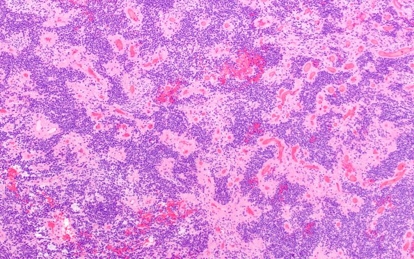
Washington University Experience | NEOPLASMS (GLIAL) | Ependymoma - Microscopic | 6A1 Ependymoma, anaplastic, RELA Fusion, WHO III (Case 6) H&E 1
Case 6 History ---- The patient was a 7-year-old girl who presented with two seizures. Brain MRI showed a 1.3 cm focal lesion within the left inferior frontal gyrus with moderate contrast enhancement and homogeneous diffusion restriction. Operative procedure: Left frontal parietal craniotomy for resection of cortical tumor. ---- 6A1,2 This is a hypercellular neoplasm with increased nuclear to cytoplasmic ratio. The tumor cells are arranged predominantly in a sheetlike architecture with prominent perivascular pseudorosettes. Mitotic activity is increased (focally up to 11/10HPF). Although scattered karyorrhectic debris is seen, necrosis is not identified. The tumor cells form a relatively well circumscribed border with adjacent brain parenchyma.

