Table of Contents
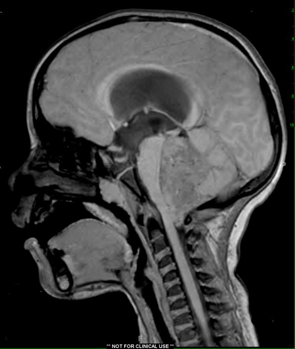
Washington University Experience | NEOPLASMS (GLIAL) | Ependymoma - Microscopic | 7A1 Ependymoma with focal anaplasia (Case 7) T1 - Copy
Case 7 History ---- The patient is a four year old male who presented with a one week history of headache and irritability, and a notable change in his balance. A CT scan demonstrated a posterior fossa mass. MRI showed a large mass lesion centered in the 4th ventricle. Clinical and radiological differential diagnosis included ependymoma and less likely, medulloblastoma, ATRT, choroid plexus papilloma, or pilocytic astrocytoma. Operative procedure: Stealth guided stereotactic suboccipital craniotomy for resection of fourth ventricular tumor. ---- 7A1,2 MRI showed a large T1-weighted (7A1) contrast enhancing (7A2) necrotic mass, centered in the 4th ventricle with calcifications and mass effect on the brainstem.

