Table of Contents
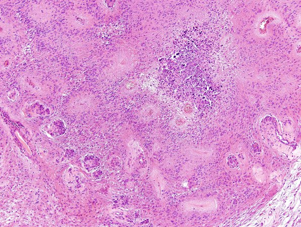
Washington University Experience | NEOPLASMS (GLIAL) | Ependymoma - Microscopic | 7B1 Ependymoma with focal anaplasia (Case 7) H&E 15
7B1-3 Sections show a tumor with multiple areas differing in appearance and cell density. Some areas show slightly increased cellularity and are composed primarily of cells with long spindled cytoplasm, and oval to elongated nuclei with finely stippled chromatin and inconspicuous nucleoli. However, scattered throughout the specimen are areas with necrosis, calcification and vascular proliferation.

