Table of Contents
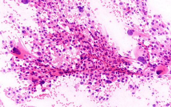
Washington University Experience | NEOPLASMS (GLIAL) | Ependymoma - Microscopic | 8A1 Ependymoma with giant cells (Case 8) H&E 5
Case 8 History ---- The patient was a 46-year-old woman who presented with a history of dizziness, paresthesia and numbness in both arms, headache, and slurred speech. MRI on 04/10 showed an irregular 2.1 x 2.1 x 3.2 cm, enhancing, T2/FLAIR hyperintense mass with internal cystic spaces, arising from the floor of the fourth ventricle, extending superiorly into the cerebral aqueduct and inferiorly to the level of the superior medulla. Susceptibility artifact was noted in the posterior lateral aspect of the tumor, likely representing calcification or blood products. Radiological differential diagnosis included ependymoma, choroid plexus papilloma, medulloblastoma, metastatic disease, and intraventricular meningioma. Operative procedure: Suboccipital craniotomy for resection of fourth ventricular tumor. ---- 8A1-5 H&E stained squash preparation performed at the time of frozen section shows numerous tumor cells, some with delicate processes attached to a small vessel, others with an epithelioid appearance as well as numerous giant pleomorphic cells (H&E)

