Table of Contents
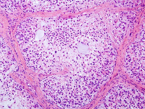
Washington University Experience | NEOPLASMS (GLIAL) | Glioblastoma, adenoid pattern | 10B3 Gliosarcoma, adenoid features (Case 10) H&E 4
10B3-5 Tumor cells are embedded in pale blue myxoid tissue that appear within a network of eosinophilic collagen-rich fibrous bands. The mucinous nodules are populated at moderate to high density by variably bipolar or epithelioid neoplastic cells with eosinophilic cytoplasm, occasional vacuoles, and small oval nuclei with variable chromatin quality. The relatively hypocellular fibrous bands are dominated by spindled cells, but show occasional areas of increased cellularity populated by cells with plump oval nuclei and mild pleomorphism. Mitotic figures are common. Vessels within the fibrous tissue show endothelial hyperplasia. The very center of the neoplasm shows bland necrosis.

