Table of Contents
Washington University Experience | NEOPLASMS (GLIAL) | Glioblastoma, adenoid pattern | 12A1 GBM, Adenoid Features (Case 12) H&E 6
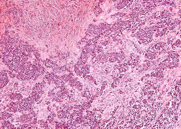
Case 12 History ---- The patient was a 64 year old woman with a prior history of melanoma. Imaging studies revealed a ring-enhancing left frontal lobe mass with a contrast enhancing satellite lesion. ---- 12A1,2 A representative section reveals a moderate to highly cellular neoplasm with both solid and focally infiltrative growth patterns. There is moderate nuclear pleomorphism and the tumor has variable morphologic features, including gemistocytic and fibrillary morphology, most prominent within the infiltrative components. Other portions of the tumor are characterized by adenoid patterns. The mitotic index is brisk and both endothelial hyperplasia and necrosis are seen focally.

